

Design of Mechanical Simulator of Artery
Senior Thesis B.Sc. Mechanical Engineering, June 2011 - May 2012
Group: Computational Instrumentation Lab, MIT
PI/Thesis Advisor: Dr. Brian Anthony
-
Goal: Design and build a mechanical simulator of an artery to test the viability of using ultrasound to make blood pressure measurements.
Previous work in the lab instrumented an ultrasound probe with load cells to measure the force applied by the probe. One potential application to measure blood pressure as an alternative to the blood pressure cuff. My project was to build a mechanical replica of the brachial artery of the arm and then use that replica to demonstrate the application viability in static and dynamic tests.
-
Constructed a mechanical model of artery and surrounding tissue,
Researched tissue stiffnesses and potential phantom materials capable of replicating those stiffnesses in tissue phantoms
Designed and constructed molds for casting tissue phantoms
Designed experiments for static and dynamic pressure tests. Collected experimental test data and summarized the results in thesis
Phantom Mechanical Design
Mechanical model of arm cross section. The artery and surrounding tissue are modeled as a stack of springs, compressed between ‘ground’ and the ultrasound probe.
On the left is the mechanical model of the arm. There are three main components: the artery surrounding muscle tissue and fluid in the artery. Their stiffnesses combine to form an overall stiffness that can be measured when the probe is compresses the structure through a known distance.
Tissue Property Requirements
Simulator artery must have similar elastic stiffness and dimensions as a real brachial artery
The tissue surrounding the artery simulator must have elastic stiffness and dimensions as the muscle surrounding the brachial artery.
Ability to view artery simulator using medical ultrasound - artery needs to be opaque to ultrasound while surrounding tissue appears transparent.
Mold Construction and Phantom Materials
The assembly included two different phantom materials, one for the artery and another for the muscle tissue.
Artery Tissue: Artery phantom was constructed of cyrogel, doped with graphite. Graphite ref and cyrogel enabled one to tune stiffness using freeze cycles. The molds are made of machinable wax, chosen for its self-releasing properties and machinability.
Muscle Tissue: ‘Muscle’ was made using co-polymer mineral oil phantom, which was transparent to the ultrasound probe. As the artery needed to be inserted into the muscle tissue phantom, the muscle phantom needed to be very durable. The co-polymer mineral oil phantom fit the bill. The mold is made of aluminum plates bolted together. Here, wax was not used as the mixture needed to be melted together and that heat would melt the wax. Additionally, the material’s durability meant that the self-release wasn’t required.
Artery cyrogel phantom in clamshell wax molds. Construction Details: Materials sourced from McMaster Carr. Wax molds made on mill. Holes are for bolts used to hold the two halves of the clamshell mold together. The number and spacing of the bolt holes were chosen so that pressure cones overlapped, eliminating leaks between mold halves.
Mineral Oil Co-polymer mixture used for muscle tissue in artery replica. Mold is made of aluminum plates bolted together. Plates were rough cut on the bandsaw and then milled to size. Bolt holes were drilled on a manual mill.
Assembly for Pulsating Flow
A diaphragm pump generates pulses of water that flow through the artery phantom. I selected the pump to mimic the volume of blood pumped with each heartbeat. The assembly includes a photologic gate to measure pulse rate. A DC motor drives the pump.
Test Results Under Static and Pulsating Flow
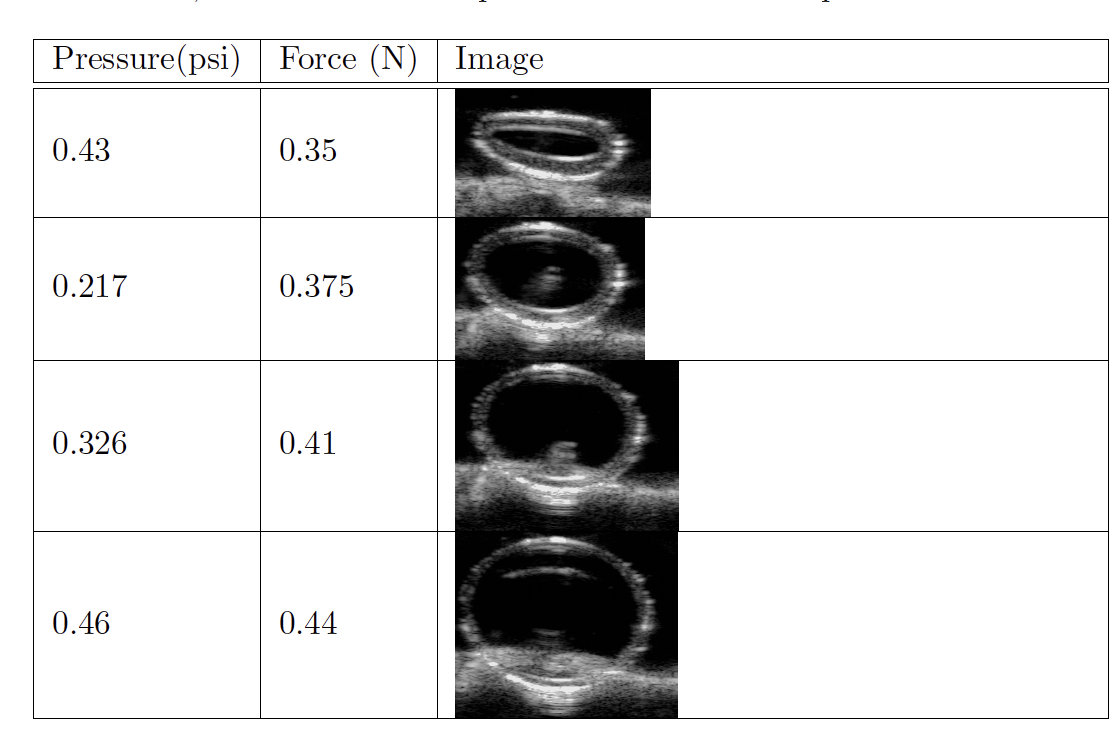
Artery under varying static fluid pressure. Instrumented ultrasound probe measured force and captured cross-section of vessel. Fluid pressure varied via chaning fluid height in connecting tube.
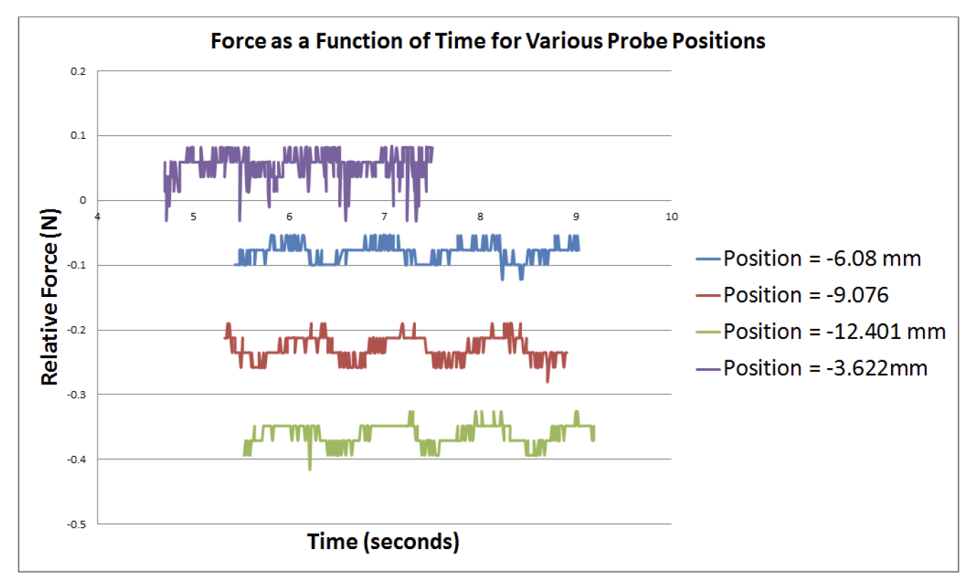
Force measurement captured by instrumented ultrasound probe, for varying probe height. Lower probe height leads to increased force magnitude, as expected.
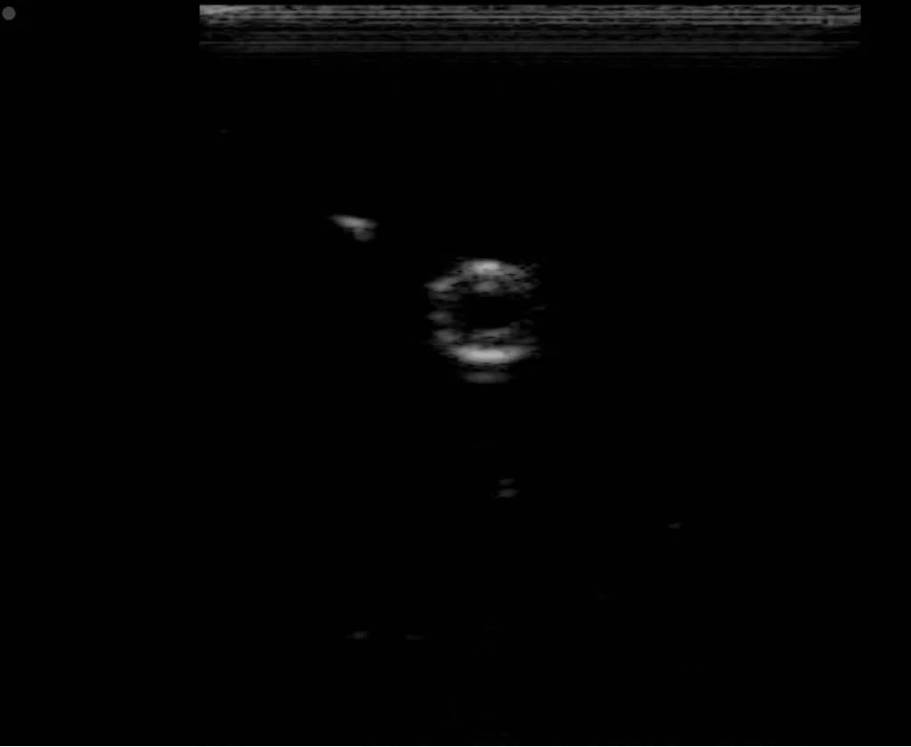
Ultrasound Images of Mechanical Simulator - Uncompressed and Uncompressed
Images from Senior Thesis
Fig 4.12 in Thesis - Uncompressed artery simulator
Fig 4.13 in Thesis - Compressed artery simulator
Fig 4.14 in Thesis - Uncompressed artery simulator, lengthwise


